Overview
We aim to contribute on future medicine by developing practical and efficient technologies and evaluation methodologies in the field of nuclear medicine (PET and SPECT) and related imaging modalities.
We aim to contribute on future medicine by developing practical and efficient technologies and evaluation methodologies in the field of nuclear medicine (PET and SPECT) and related imaging modalities.
2018.5.23 Uploded Dr. Shdahara's talk at Tohoku university science cafe
2018.4.1 Information of admissions is here.
Department of Quantum Science and Energy Engineering
6-6-01-2, Aramaki Aza Aoba Aoba-ku, Sendai, Miyagi 980-8579, Japan
E-mail: miho.shidahara.c2@tohoku.ac.jp
TEL�F+81-22-795-7928
Research Building-M.A.E.
Asscoiate Professor

Research fellow

D2

M2(DDprogram, EC-Lyon)

M2

M2

M1

M1

M1

B4

B4
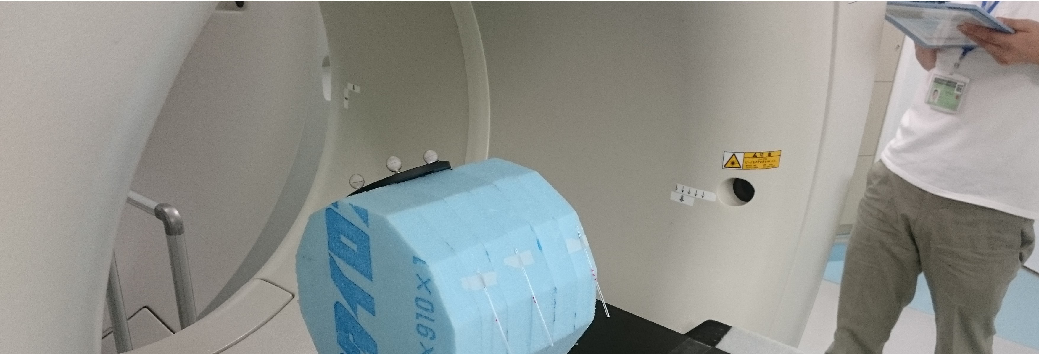
Quantification of functional imaging can be degraded by limited spatial resolution, incomplete processing, subject�fs motions and so on. To improve the quantification, we are developing methodologies for motion correction and image-processing.
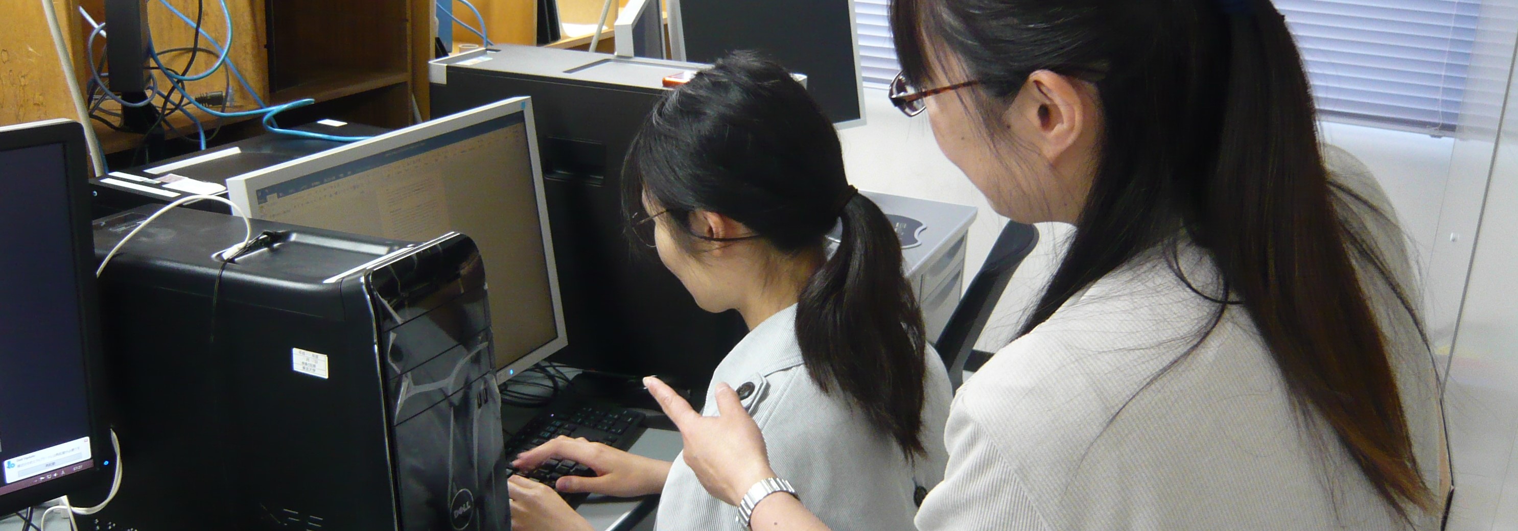
Computer-aided virtual clinical trial is a kind of simulation techniques for human body but its application have great potential as before having clinical trial in many ways. We try to develop the system for efficient development of radioligands.
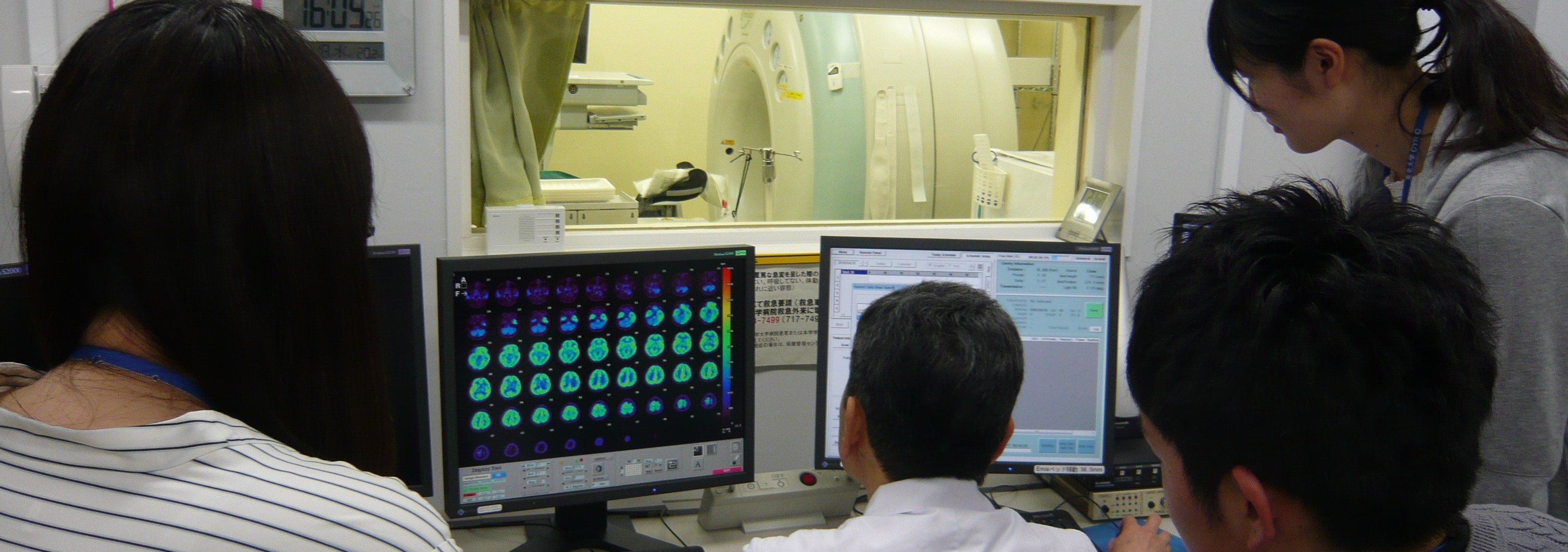
Lesion-detection ability is one of important diagnostic factors for medical imaging. We try to predict the impact of introducing new technologies by numerical observer model.
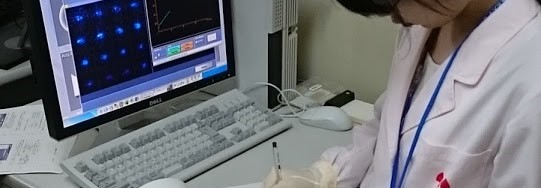
Internal radiation exposure in nuclear medicine is inevitable and have to be estimated for risk-benefit management. Human radiation dose suffered from nuclear medicine can be estimated from time-series measurement of the biodistribution of the injected radioligand. We aim to develop noninvasive methodologies further practical applications.